| Humoralpathologisches Forschungslabor | HLB blood test
HLB blood test
The HLB blood test offers a humoral pathological diagnostics which provides a preventive medical insight in the total metabolism.
The abbreviation HLB stands for Dr Heinz Heitan, Dr Philippe de LaGarde och Dr H Leonard Bolen who by experimenting developed this specific examination of blood from the 1930ies to the 1950ies.
The German physician Dr Heinz Heitan, who lived in exile in Nice, brought to attention the test during the 1950ies and reported this in the cancer conference in Vienna in 1954. He referred to the blood test in conjunction with the micro organisms in blood described by Prof. Günther Enderlein and Dr Wilhelm von Brehmer who both thought that these were responsible for the genesis of cancer.
How does the HLB test work?
One drop of dried capillary blood is examined with a simple optical microscope. When the blood dries erythrocyte cluster and fibrin net are formed and the sedimentation pattern which appears is then evaluated.
Contrasting to the common quantitative blood examinations which are based on the amount of substances contained in blood, the HLB blood test provides a qualitative blood analysis which enables drawing conclusions regarding the total functional state of the blood.
Originally, the American surgeon H. Leonard Bolen intended to use the sample as a screening test for early diagnostics of gastrointestinal tumours. The test was used by multiple physicians in a large number of cases. It was then evident that the test did not allow specific cancer diagnoses, since there were also positive results for several other diseases.
However, from the blood test conclusions could be drawn about the general state of blood. Specific morphological- and colour changes of the dried blood drops indicate changes in the metabolism of the organs, circulation and the state of the red blood cells.
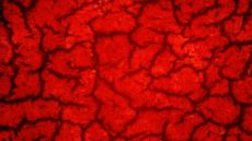 |
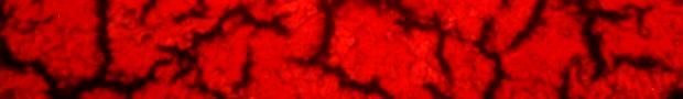
| Picture 1: Healthy blood |
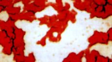 |
| Picture 2: Unhealthy blood |
Healthy blood spreads evenly on the slide and has a homogenous structure when it has dried, see picture 1. The pattern reminds of the macroscopic structure of cerebrum. The black coloured reticular threads are formed by the fibrin. The red spots consist of the clotted erythrocytes.
The HLB test reveals an attack on various organ systems as it differs from the healthy blood structure. This occurs already before these attacks have been clinically manifested or by diverging laboratory parameter results.
Picture 2 shows changes in the blood structure in unhealthy blood. The red blood cells are diminished and partly destroyed. The structure appears to be ”torn” and the fibrin net almost dissolved. In the empty space between the clusters of erythrocytes there are isolated blood cells and clusters of very small granules. Haemolysis has occurred. In the old humoral pathological language this would have been referred to as ”decomposition of blood”.
What is the HLB blood test telling us?
With the HLB test it is possible to determine 5 phases in the blood deterioration which, supported by Enderlein, are described as endobiosis stages.
With endobiosis Enderlein described a total complex of chronic and degenerative diseases including cancer. According to his research, a number of diseases of which the aetiology until now is unknown to the faculty medicine, are caused by one single endogenous polymorphic micro organism in blood. Due to its smallness this micro organism has generally been overlooked by the haematologists. As a zoologist, Enderlein regarded this micro organism as the ”primary symbiont” of the mammals and gave it the name ”endobiont”.
The endobionts appear in the organism in different live phases and forms of development. During life and due to a number of toxic influences, the originally harmless endobionts may develop disease causing characteristics. Enderlein wrote:
”These micro organisms – by me called Endobionts – are harmless in their primitive form, multiply through their life to an astronomically large number, develop further when humans live in a nature-adverse-lifestyle to higher valence forms and form colonies living on nearby erythrocytes, causing a clotting of the blood – thrombosis. This causes disturbances in all body organs which could result in a complete paralysis of the functions in the respective organ. In this way various diseases arise caused by the same process – the development from the primitive form into the higher valence form”. [cit. n. E. Krämer: Leben und Werk von Prof. Dr. phil. Günther Enderlein (1872-1968). Med. Diss. Frankfurt/M. 2006 S.235]
Humoral pathological basics
In his papers, Enderlein referred to research by the pathologist from Vienna, Prof. Carl von Rokitansky (1804-1878), who was one of the last great representatives for humoral pathology during the 18th century.
Rokitansky opposed the new medical thesis of Rudolf Virchow, cell pathology. Virchow had in general placed the disease processes on a cellular level and thereby replaced the old humoral pathological medical concept.
Contrary to this, Rokitansky claimed that formation of fibrin caused a number of diseases, where he could distinguish a biological process.
Rokitansky meant that the pathological formation of fibrin was caused by a number of chronic, degenerative diseases which in an inflammatory process would lead to a restructuring of connective tissue and loss of function in the organs.
Enderlein, again, established the connection between the fibrin and the endobionts in blood. He described the fibrin as a primitive growth phase in the endobionts.
It may sound remarkable, but Enderlein’s conclusion is confirmed by the imposing French chemist and medical professor Pierre Jacques Antoine Béchamp (1816-1908) in his research on blood coagulation.
Béchamp could show that the fermentation process in the organism, including the fibrin formation, purely could be considered as disturbances in the metabolism among lower micro organisms. He separated living from dead ferments (enzymes). The living enzymes he named “microzymas”. They were to be the producers of the lifeless enzymes.
Béchamp also isolated the living enzymes from blood and various human tissues. He could show that microzymas provoked pathological changes in the cells during diseases which finally led to the destruction of those.
In his monograph “The Blood and it’s Third Anatomical Element”, Béchamp pointed out the microzymatical properties of fibrin and its evolution to a bacterial life form.
HLB test versus diagnosis of vital blood in darkfield
These microbial courses of events in blood become visible through microscopic examination of healthy blood in darkfield.
“The vital blood diagnostics in darkfield according to Enderlein”, has been implemented in our laboratory for years. These particular examinations of blood were part of a comprehensive blood diagnostics which Enderlein effectuated in his humoral pathological research laboratory (IBICA). The method is very time consuming and does not at all allow for drawing the multitude of conclusions, which blood diagnosticians claim possible. As a consequence Enderlein wrote as follows:
“It can naturally not be expected that the examination enables a firm diagnosis ( e.g. cancer); it purely allows for summarizing a preparedness for cancer. This question is superfluous when endobiosis characteristics and concepts have been made clear. Cancer as such, only lets itself be diagnosed in connection with histological and clinical findings. The examination focuses amongst other things on establishing the strength of the attack by the erythrocytes, the core of the leukocyte and the leukocyte plasma and simultaneously the valence.” [G. Enderlein:IBICA-Information. Juni 1954]
The HLB test is compared to the darkfield diagnostics less spectacular, but facilitates the evaluation of the deterioration of blood (endobiosis).